


Your cart is empty
This quantity of item cannot be added to the cart
Continue shopping
Health hub
Learn everything you need to know about a healthy and balanced life in one place. From your cholesterol levels and liver function to women's health, we cover a range of topics to help improve your health knowledge.
Mens Health
View all-

Best Multivitamins For Men
Multivitamins remain one of the most widely used supplements among men in the UK. Busy schedules, inconsistent diets, training demands, stress, and reduced sunlight exposure can all contribute to micronutrient...
Best Multivitamins For Men
Multivitamins remain one of the most widely used supplements among men in the UK. Busy schedules, inconsistent diets, training demands, stress, and reduced sunlight exposure can all contribute to micronutrient...
-

11 Best Men’s Hair Products of 2026
Selection of the best products for Men's Hair requires navigation of a lot of hair sprays, waxes, gels, and pomades, and all these products claim to be the best ones....
11 Best Men’s Hair Products of 2026
Selection of the best products for Men's Hair requires navigation of a lot of hair sprays, waxes, gels, and pomades, and all these products claim to be the best ones....
-

10 Best Natural Testosterone Supplements
Testosterone, a hormone mostly produced in the testicles for men and the ovaries and adrenal glands for women, has a significant impact on our health. It is responsible for maintaining...
10 Best Natural Testosterone Supplements
Testosterone, a hormone mostly produced in the testicles for men and the ovaries and adrenal glands for women, has a significant impact on our health. It is responsible for maintaining...
Women's Health
View all-

Best Multivitamins For Women
Dr Muhammad Zeeshan AfzalModern women's health needs are rarely about one nutrient at a time. In the UK, busy schedules, inconsistent meal patterns, limited daylight exposure, dietary choices such as vegetarian or vegan...
Best Multivitamins For Women
Dr Muhammad Zeeshan AfzalModern women's health needs are rarely about one nutrient at a time. In the UK, busy schedules, inconsistent meal patterns, limited daylight exposure, dietary choices such as vegetarian or vegan...
-

Best Panty Liners for Comfort in 2026, Tried an...
Dr Muhammad Zeeshan AfzalThe right pantyliner makes a big difference in hygiene and everyday comfort. Women who are concerned about their comfort during light flow days choose them. In 2026, many market options...
Best Panty Liners for Comfort in 2026, Tried an...
Dr Muhammad Zeeshan AfzalThe right pantyliner makes a big difference in hygiene and everyday comfort. Women who are concerned about their comfort during light flow days choose them. In 2026, many market options...
-

11 Best Shaving Creams for Women
Dr Muhammad Zeeshan AfzalShaving is vital to many women's grooming routines, and the choice of the right shaving creams makes a huge difference. A good shaving cream not only ensures a closer shave...
11 Best Shaving Creams for Women
Dr Muhammad Zeeshan AfzalShaving is vital to many women's grooming routines, and the choice of the right shaving creams makes a huge difference. A good shaving cream not only ensures a closer shave...
Pet Health
View all-

Best Joint Supplements for Dogs UK
Joint supplements can be a smart part of a dog’s long-term mobility plan—especially for older dogs, large breeds, dogs with early stiffness, or those recovering from heavy activity. Canine osteoarthritis...
Best Joint Supplements for Dogs UK
Joint supplements can be a smart part of a dog’s long-term mobility plan—especially for older dogs, large breeds, dogs with early stiffness, or those recovering from heavy activity. Canine osteoarthritis...
-

Best Diarrhea Medicine for Dogs of 2026
Dog Diarrhoea Relief: Why is it Important? Diarrhoea in dogs isn’t a particular disease but a symptom of a lot of possible health problems. In dogs with diarrhoea, the food...
Best Diarrhea Medicine for Dogs of 2026
Dog Diarrhoea Relief: Why is it Important? Diarrhoea in dogs isn’t a particular disease but a symptom of a lot of possible health problems. In dogs with diarrhoea, the food...
-

10 Best Dog Multivitamins for Your Furry Friend
Pet supplements are a vital part of taking proper care of your pet since they will help to complement a balanced diet. Dogs also require certain nutrients for them to...
10 Best Dog Multivitamins for Your Furry Friend
Pet supplements are a vital part of taking proper care of your pet since they will help to complement a balanced diet. Dogs also require certain nutrients for them to...
Bone & Joint Health
View all-

Best Supplements for Joint Pain Relief
Joint pain is one of the most common musculoskeletal complaints in the UK, affecting adults across all age groups and activity levels. In clinical practice, Dr Zeeshan Afzal frequently sees...
Best Supplements for Joint Pain Relief
Joint pain is one of the most common musculoskeletal complaints in the UK, affecting adults across all age groups and activity levels. In clinical practice, Dr Zeeshan Afzal frequently sees...
-

Best Calcium Supplements in UK
Calcium is an essential mineral required for normal bone structure, muscle contraction, nerve signalling, and blood clotting. While calcium is best obtained through food, many people in the UK consider...
Best Calcium Supplements in UK
Calcium is an essential mineral required for normal bone structure, muscle contraction, nerve signalling, and blood clotting. While calcium is best obtained through food, many people in the UK consider...
-

Crepitus & Clicking Joints: Symptoms, Treatment...
Crepitus, or joint popping, is the term for audible noises produced by a moving joint, such as popping, cracking, and grinding. When these "pops" occur, pressure seems to be released...
Crepitus & Clicking Joints: Symptoms, Treatment...
Crepitus, or joint popping, is the term for audible noises produced by a moving joint, such as popping, cracking, and grinding. When these "pops" occur, pressure seems to be released...
Sports Performance
View all-

Best Huel Flavours Reviewed: What’s Worth Buying
Meal replacement products have become increasingly common in the UK, particularly among individuals seeking a practical way to maintain nutritional consistency alongside demanding schedules. One of the most recognisable brands...
Best Huel Flavours Reviewed: What’s Worth Buying
Meal replacement products have become increasingly common in the UK, particularly among individuals seeking a practical way to maintain nutritional consistency alongside demanding schedules. One of the most recognisable brands...
-

Best MyProtein Flavours in 2026
Protein powders are among the most widely used sports nutrition products in the UK, extending well beyond bodybuilding into general health, weight management, and everyday nutrition. Within this landscape, MyProtein has...
Best MyProtein Flavours in 2026
Protein powders are among the most widely used sports nutrition products in the UK, extending well beyond bodybuilding into general health, weight management, and everyday nutrition. Within this landscape, MyProtein has...
-

The Best Protein Waters of 2026
Protein waters have become one of the biggest innovations in modern sports nutrition, offering a refreshing way to stay hydrated while increasing daily protein intake. Over the past year, protein...
The Best Protein Waters of 2026
Protein waters have become one of the biggest innovations in modern sports nutrition, offering a refreshing way to stay hydrated while increasing daily protein intake. Over the past year, protein...
Vitamin & Nutrition Blog
View all-

Best Vitamin D3 and K2 Drops
By Dr Muhammad Zeeshan Afzal, MBBS | FCPS | MRCP A comprehensive editorial guide. Last updated November 2026. Quick answer: What are the best vitamin D3 and K2 drops in...
Best Vitamin D3 and K2 Drops
By Dr Muhammad Zeeshan Afzal, MBBS | FCPS | MRCP A comprehensive editorial guide. Last updated November 2026. Quick answer: What are the best vitamin D3 and K2 drops in...
-

The Best TMG Supplements Ranked
By Dr Muhammad Zeeshan Afzal Last clinically reviewed: 7 May 2026 | Reading time: 38 minutes | Medically reviewed by Welzo's clinical team The best TMG (trimethylglycine) supplement in 2026...
The Best TMG Supplements Ranked
By Dr Muhammad Zeeshan Afzal Last clinically reviewed: 7 May 2026 | Reading time: 38 minutes | Medically reviewed by Welzo's clinical team The best TMG (trimethylglycine) supplement in 2026...
-

4 Best Apigenin Supplements of 2026
Apigenin is a naturally occurring plant flavonoid found in chamomile, parsley, celery, oregano, thyme, and several other herbs and vegetables. It is best known for its potential calming, antioxidant, and...
4 Best Apigenin Supplements of 2026
Apigenin is a naturally occurring plant flavonoid found in chamomile, parsley, celery, oregano, thyme, and several other herbs and vegetables. It is best known for its potential calming, antioxidant, and...
Blood Tests
View all-

Best Home Blood Tests in the UK, Ranked by a Do...
Blen TesfuHome blood test kits have become one of the most practical ways for people in the UK to understand their health without long NHS wait times or private clinic appointments....
Best Home Blood Tests in the UK, Ranked by a Do...
Blen TesfuHome blood test kits have become one of the most practical ways for people in the UK to understand their health without long NHS wait times or private clinic appointments....
-

Introducing BloodTestsOnline.co.uk – The UK's N...
Dr Adam WilliamsWe are proud to announce the launch of BloodTestsOnline.co.uk, the UK's first fully dedicated home blood testing platform. Built to address the growing demand for fast, accurate, and convenient diagnostic...
Introducing BloodTestsOnline.co.uk – The UK's N...
Dr Adam WilliamsWe are proud to announce the launch of BloodTestsOnline.co.uk, the UK's first fully dedicated home blood testing platform. Built to address the growing demand for fast, accurate, and convenient diagnostic...
-

What Level of Eosinophils Indicate Cancer?
Dr Sadia SaeedEosinophils are a type of white blood cell that make up a tiny portion of the white blood cells in the bloodstream. Like other white blood cells, it also plays...
What Level of Eosinophils Indicate Cancer?
Dr Sadia SaeedEosinophils are a type of white blood cell that make up a tiny portion of the white blood cells in the bloodstream. Like other white blood cells, it also plays...
Antibiotics
View all-

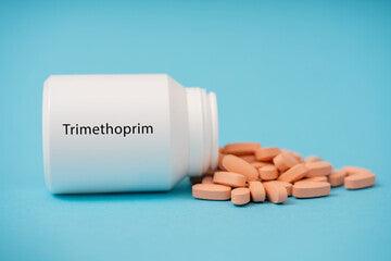
Trimethoprim vs. Other Antibiotics: A Comparati...
Antibiotics are potent drugs employed in modern medicine that kill or inhibit the growth of microorganisms, specifically bacteria, and certain parasites, to prevent and treat infections in humans and animals...
Trimethoprim vs. Other Antibiotics: A Comparati...
Antibiotics are potent drugs employed in modern medicine that kill or inhibit the growth of microorganisms, specifically bacteria, and certain parasites, to prevent and treat infections in humans and animals...
-

The Future of Antibiotics: Innovations and Chal...
The Future of Antibiotics: Innovations and Challenges Antibiotics, often hailed as one of the greatest medical discoveries of the 20th century, have revolutionized the field of medicine and saved countless...
The Future of Antibiotics: Innovations and Chal...
The Future of Antibiotics: Innovations and Challenges Antibiotics, often hailed as one of the greatest medical discoveries of the 20th century, have revolutionized the field of medicine and saved countless...
-

Antibiotics for Dental Issues: A Closer Look at...
Antibiotics for Dental Issues: A Closer Look at Oral Health Antibiotics have played a pivotal role in modern medicine, revolutionizing the treatment of bacterial infections and saving countless lives. While...
Antibiotics for Dental Issues: A Closer Look at...
Antibiotics for Dental Issues: A Closer Look at Oral Health Antibiotics have played a pivotal role in modern medicine, revolutionizing the treatment of bacterial infections and saving countless lives. While...
Beauty & Cosmetics
View all-

Best Women’s Perfumes in 2026, According to Fra...
Perfume is not just a fragrance but a way of expressing yourself and finishing with a touch that makes a lasting impression. In 2026, the world of women's fragrances is...
Best Women’s Perfumes in 2026, According to Fra...
Perfume is not just a fragrance but a way of expressing yourself and finishing with a touch that makes a lasting impression. In 2026, the world of women's fragrances is...
-

Top 10 Men’s Perfumes of 2026, Recommended by E...
Choosing an excellent perfume is not just about choosing a scent but about leaving a lasting impression throughout the day. Men's perfumes are different from home perfumes and women's perfumes...
Top 10 Men’s Perfumes of 2026, Recommended by E...
Choosing an excellent perfume is not just about choosing a scent but about leaving a lasting impression throughout the day. Men's perfumes are different from home perfumes and women's perfumes...
-

Top 10 Men’s Hair Pomades of 2026
Top 10 Men's Hair Pomades of 2026 The selection of an ideal pomade makes a big difference in hairstyle, whether looking for a more textured or voluminous finish or a...
Top 10 Men’s Hair Pomades of 2026
Top 10 Men's Hair Pomades of 2026 The selection of an ideal pomade makes a big difference in hairstyle, whether looking for a more textured or voluminous finish or a...
Baby & Child
View all-

Best Liquid Vitamins For An Autistic Child
Liquid vitamins can be a practical option for autistic children when tablets are a non-starter. Sensory sensitivities, swallowing difficulties, selective eating, and routine rigidity can all make standard children's supplements...
Best Liquid Vitamins For An Autistic Child
Liquid vitamins can be a practical option for autistic children when tablets are a non-starter. Sensory sensitivities, swallowing difficulties, selective eating, and routine rigidity can all make standard children's supplements...
-

10 Best Nasal Aspirators of 2026
Choosing the right nasal aspirator for both babies and adults can make a huge difference - especially during cold and allergy seasons. Nasal congestion impacts sleep, feeding, and overall comfort,...
10 Best Nasal Aspirators of 2026
Choosing the right nasal aspirator for both babies and adults can make a huge difference - especially during cold and allergy seasons. Nasal congestion impacts sleep, feeding, and overall comfort,...
-

Baby Acne and Breast Milk: Causes and Treatment
Many new parents are surprised to discover small red or white bumps on their baby's face, backs, cheeks, and chin, often appearing weeks or months after birth. This common condition,...
Baby Acne and Breast Milk: Causes and Treatment
Many new parents are surprised to discover small red or white bumps on their baby's face, backs, cheeks, and chin, often appearing weeks or months after birth. This common condition,...
Cold & Flu
View all-

11 Best Cough Sweets to Relieve Sore Throat
What is a Sore Throat? Painful, scratchy, or irritated sensation in all or part of the throat, especially with difficulty swallowing. A sore throat can range from just a minor...
11 Best Cough Sweets to Relieve Sore Throat
What is a Sore Throat? Painful, scratchy, or irritated sensation in all or part of the throat, especially with difficulty swallowing. A sore throat can range from just a minor...
-

12 Best Teas for Sore Throat to Help You Get Th...
Cold and flu season is here again, bringing along stuffy noses and sore throats. It can feel like a never-ending cycle of discomfort, but thankfully, tea can help you feel...
12 Best Teas for Sore Throat to Help You Get Th...
Cold and flu season is here again, bringing along stuffy noses and sore throats. It can feel like a never-ending cycle of discomfort, but thankfully, tea can help you feel...
-

Why does putting vicks on your feet stop Coughing?
Vicks VapoRub, a ubiquitous name in households worldwide, stands as a testament to the enduring legacy of over-the-counter remedies. Its formulation, a blend of camphor, eucalyptus oil, and menthol, has...
Why does putting vicks on your feet stop Coughing?
Vicks VapoRub, a ubiquitous name in households worldwide, stands as a testament to the enduring legacy of over-the-counter remedies. Its formulation, a blend of camphor, eucalyptus oil, and menthol, has...
Erectile Dysfunction
View all-

Erectile Dysfunction: Symptoms, Causes, Diagnos...
A Comprehensive Guide to Erectile Dysfunction: Symptoms, Causes, Diagnosis and Treatment Erectile Dysfunction (ED), also known as impotence, is a common male health issue in which a male is either...
Erectile Dysfunction: Symptoms, Causes, Diagnos...
A Comprehensive Guide to Erectile Dysfunction: Symptoms, Causes, Diagnosis and Treatment Erectile Dysfunction (ED), also known as impotence, is a common male health issue in which a male is either...
-

Simple Trick to Cure ED: 7 Simple Steps
7 Simple Tricks to Cure Erectile Dysfunction Explanation of Erectile Dysfunction (ED) Erectile dysfunction (ED) is a prevalent health issue affecting millions of men worldwide. It is a condition characterised...
Simple Trick to Cure ED: 7 Simple Steps
7 Simple Tricks to Cure Erectile Dysfunction Explanation of Erectile Dysfunction (ED) Erectile dysfunction (ED) is a prevalent health issue affecting millions of men worldwide. It is a condition characterised...
-

Can a Blood Test Check for Erectile Dysfunction?
Also known as impotence, erectile dysfunction (ED) is a common condition affecting millions of men worldwide. It refers to the inability to maintain or achieve an erection sufficient for sexual...
Can a Blood Test Check for Erectile Dysfunction?
Also known as impotence, erectile dysfunction (ED) is a common condition affecting millions of men worldwide. It refers to the inability to maintain or achieve an erection sufficient for sexual...
Hair Loss
View all-

7 Best HRT for Hair Loss: Reviewed by Experts
Dr Muhammad Zeeshan AfzalHair loss is a common issue regardless of gender and is mostly linked to hormonal imbalances and fluctuations. Other factors, such as environmental factors, genes, and lifestyle, have some role,...
7 Best HRT for Hair Loss: Reviewed by Experts
Dr Muhammad Zeeshan AfzalHair loss is a common issue regardless of gender and is mostly linked to hormonal imbalances and fluctuations. Other factors, such as environmental factors, genes, and lifestyle, have some role,...
-

Double Crown Hair: What Is It and What Does It ...
Dr Sadia SaeedMost people have a certain pattern of hair development near the head's crown. The head starts to bend downward towards the rear of the skull at this point. A normal...
Double Crown Hair: What Is It and What Does It ...
Dr Sadia SaeedMost people have a certain pattern of hair development near the head's crown. The head starts to bend downward towards the rear of the skull at this point. A normal...
-

Topical Finasteride: Does It Work for Hair Loss?
Dr Sadia SaeedHair loss is a common problem that affects millions of people all over the world. It has many different causes that range from non-modifiable causes such as age and genetics...
Topical Finasteride: Does It Work for Hair Loss?
Dr Sadia SaeedHair loss is a common problem that affects millions of people all over the world. It has many different causes that range from non-modifiable causes such as age and genetics...
 Rated Excellent by 26,523+ Reviews